Case study – ocular fracture
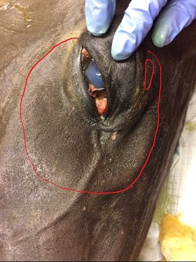
This was a recent case. The horse had been kicked by another horse and a depression involving a good part of the orbit was evident (outlined here in red in the first photo).
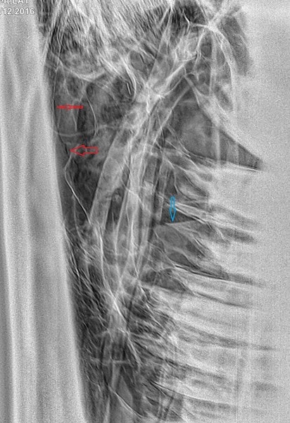
Radiographs were taken, the teeth look OK and there are various fluid lines within the sinuses from the blood (second photo: blue arrows are fluid lines and red arrows are at either end of a fracture line).
There was slight bulging of the eyeball with partial prolapse of the third eyelid with a mild purulent discharge. Staining of the cornea revealed a shallow but extensive corneal ulcer covering the ventral half of the globe which was from a sharp edge of fractured bone on the ventral orbit. Palpation of the orbit revealed disruption to over half of the circumference of the orbit.
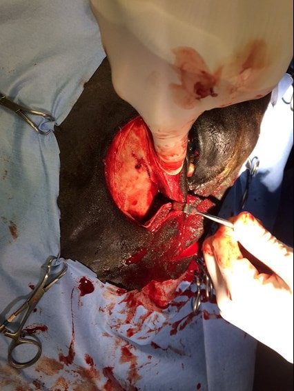
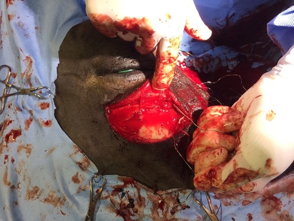
An initial attempt was made to elevate the dorsal and ventral fragments using elevators and Steinman pins through stab incisions. It became clear that this would not produce the leverage required and so two semi-circular incisions were made dorsally and ventrally but not connected to preserve the nasolacrimal duct (assuming it had not already been severed by a bone fragment). Exposure of the fracture showed that there were at least eight large fragments with several smaller pieces of loose bone being discarded.
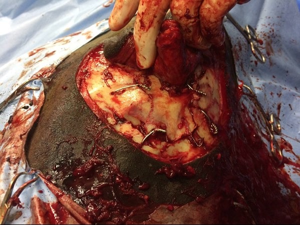
Some fragments were depressed to the extent that they were coursing vertically into the head, perpendicular to their normal position. The fractures extended around the dorsal margin of the orbit. The fragments were gradually levered back into their normal position but it also was clear that they would not be self-retaining and so adjacent fragments were secured to one another using stainless steel wire. One large central loose fragment was obscured by the tissue between the two incisions and so the incisions were connected rostrally.
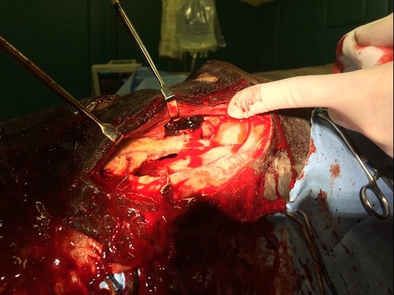
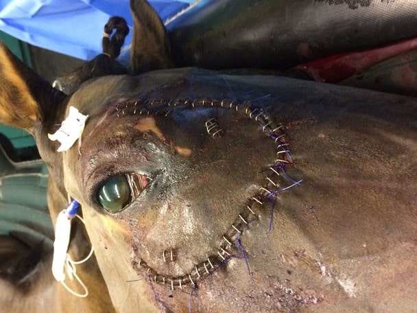
This then allowed anatomical reconstruction using five wires in total. The orbit was largely reconstructed although a defect remained at the medial canthus through bone loss but the orbit contacting the globe was confirmed by palpation to be smooth. At the end of surgery, a more normal contour to the face could be appreciated. The prolapse of the third eyelid had also resolved. Because of the severity of the corneal ulceration and the delicacy of the area around the eye, a subpalpebral lavage system was placed to allow remote medication of the eye.
So the bottom line to this case is that radiographs will almost certainly underestimate the damage, and while a CT would be great, access to a CT or finances may not always be in place.
Ultrasonography is probably much more useful – I didn’t ultrasound this cases because of finances, but in retrospect if I had I would be had a better idea of how many pieces there were. Or as we described it to the owner, it looked like a rock had been thrown through a plate glass window.
So in this case the integrity of the eyeball and adjacent structures was the main concern along with reconstructing the orbit, but in more non-critical areas, a more conservative approach can be taken.
