Doppler ultrasonography in equine reproduction
A passage explaining the usefulness of color and spectral Doppler in clinical situations involving equine ultrasonography.
Doppler ultrasonography in equine reproduction
B-mode ultrasonography was first described in equine stud work in 1980, and since then both techniques and technologies have advanced almost unrecognizable. Modern ultrasound machines are portable and powerful, giving vastly superior image quality compared to much bulkier and more expensive machines of even 5 years ago. Similarly research has ensured that our understanding of what we are able to see with the newer equipment is leading to a much improved ability to manipulate both natural and artificial breeding programs to increase success.
Early work concentrated on the 2D grey scale image known as B-mode (brightness mode) and this still constitutes the major role of ultrasonography in equine stud practice; however, the increasingly widespread availability of both color and spectral Doppler capabilities has led to an interest in their potential applications in this exciting area of equine practice.
Color Doppler is very useful in all species and in many clinical situations for giving an ‘overview’ of blood flow – its presence or absence, direction, and a semi-quantitative assessment of flow velocity. It can be useful to facilitate anatomical orientation such as in making measurements of the combined thickness of the uterus and placenta (CTUP) as a means of assessing placental health and the possible development of placentitis in mid-late gestation mares. It can be useful in assessing the likely nature of certain ovarian structures such as haemorrhagic anovulatory follicles (HAFs), and changes in color Doppler signal in pre-ovulatory follicles have been shown to drop off in the four hours prior to ovulation. It can also be applied in the transition period to assist decision-making about the likely ovulatory or anovulatory nature of dominant follicles.
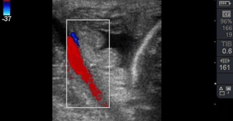
Fig. 1 Color Doppler image of the uterine artery used to ensure correct transducer placement for taking measurements of the combined thickness of the uterus and placenta (CTUP). Image courtesy of James Crabtree BVM&S CertEM(StudMed) MRCVS, Equine Reproductive Services (UK) Ltd.
There is also research being carried out to assess the usefulness of spectral Doppler, for example for assessing how uterine artery blood flow (velocity, flow volume, and resistance indices) might relate to fetal growth and development, although at this stage more work is needed.
The use of Doppler ultrasound in equine stud work is not confined to mares either; assessment of resistance indices has been shown to be useful as an indicator of testicular dysfunction in stallions.This is an exciting area of development for equine stud practice and no doubt continuing technological advancements and scientific research will lead to a better understanding of the subject, further cementing the place of ultrasonography as an indispensable tool for the equine stud practitioner and hopefully leading to ever-improving results for the breeding industry and improved horse welfare.