Equine case study – worse than a kick in the teeth
This is an older case but I was reminded by a friend about it last week. It also demonstrates how far radiographic image quality has come in ten years.
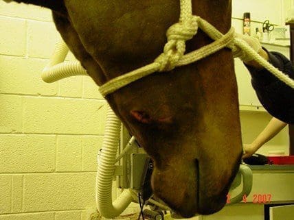
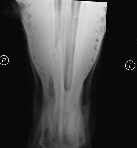
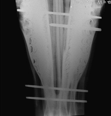
The above horse was first seen in the March for a wound on his right mandible, facial swelling, and an inability to eat. Radiographs showed a unilateral displaced fracture of the right mandibular body between the first and second cheek teeth. There was a 4 cm x 2 cm foul smelling wound on his right lateral mandible in the region of the fracture.
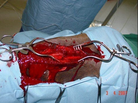
Under general anaesthesia and in dorsal recumbency, the fracture was exposed via a 10cm ventral incision centred over the fracture. Necrotic tissue, food, and haematomas were removed from the fracture gap using bone curettes and a Volkmann spoon. It was found that the fracture had split the first cheek tooth obliquely and that the wound on the face communicated with the fracture site and also to a mucosal wound within the mouth. The first cheek tooth was not removed at the time of the fracture repair because the tooth would add stability to the repair. The fracture passed obliquely across the ramus. Bone reduction forceps were applied and good fracture reduction was achieved.
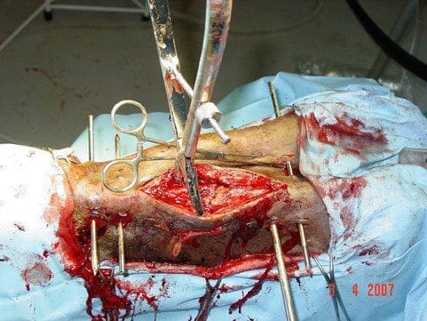
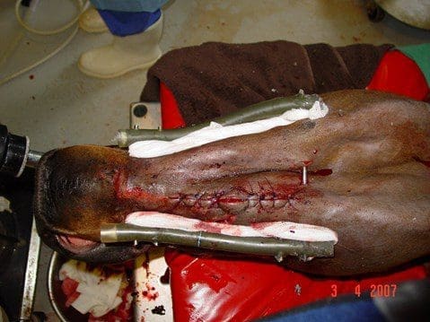
An external fixator consisting of four bone pins passed through the mandible bilaterally was applied. Two pins were on either side of the fracture. The fixator consisted of three 4 mm pins and one 5mm pin. The 5mm pin was the third pin caudally and could only be inserted through the right ramus and just into the left ramus but without passing completely through the ramus. Fracture reduction was confirmed and the pins were cut with bolt cutters 4 cm from the skin. Two lengths of tubing (a disused nasogastric tube) were cut to a length that would span all the pins with 2cm more at either end. The ends were plugged with swabs and then the tubing filled with Technovit. Once the Technovit had cured, the fracture reduction forceps were removed and the fracture was held firmly in reduction. The ventral incision was closed with 2-0 PDS in the SQ and 0 Prolene in the skin. A 2cm area in the middle of the incision was left open for drainage. The wound on the face was gently debrided with a Volkmann spoon. The pin/skin interfaces were covered with swabs soaked in intramammary antibiotics. Radiographs were taken on numerous occasions and were inconclusive with regard to tooth root involvement.
The horse ate willingly on return to his box. Antibiotic therapy was continued and well as administration of phenylbutazone. The pins were left in place for five weeks and then removed.
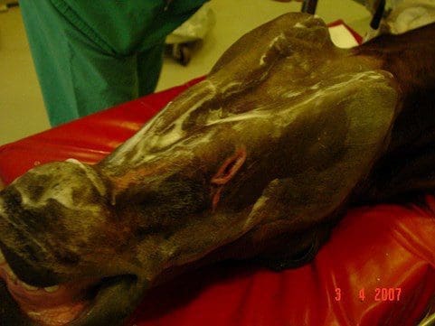
On readmission to the hospital in the August, there was purulent discharge coming from the lateral aspect of the right mandible in the area of the original wound. The incision on the ventral aspect of the mandible via which the fracture was repaired had healed well and no communication with the mouth was evident. The draining tract was curetted under a short general anaesthetic but this failed to resolve the problem.
Shortly therafter the daily wound flushing showed that saline was now exiting from the mouth. An oral examination showed that the fractured first cheek tooth had displaced further and confirmed its involvement in the persistent infection. (Yes, I know a CT would have been useful…)
Therefore, under general anaesthesia and a buccotomy and alveolar osteotomy was performed on the right mandible. The fractured first cheek tooth was removed. During surgery it was noted that the infected tract lead to the pulp cavity of the second cheek tooth and consequently this tooth was removed as well. The empty tooth sockets were filled with bandage impregnated with BIPP and a false crown made with polymethamethacrylate to protect the area from the mouth. The buccotomy incision was closed in multiple layers and beginning five days after surgery, the bandage material was removed in small portions twice daily. The surgical incision healed by primary intention and the staples were removed two weeks after surgery. The dental impression compound was checked daily and the last portion of bandage was removed on the 19th September. The original wound sealed over within two days of the last of the bandage being pulled out. The gelding did well and entered race training.
Looking at the Racing Post website, the horse went on to race and died as an eleven year old.
