Equine ocular ultrasonography
A case study explaining the Indications, preparation and techniques required to effectively perform diagnostic equine ocular ultrasonography.
Indications
Any situation in which the view into the globe is obscured. For example:
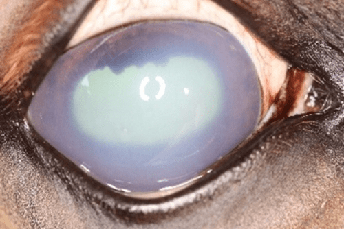
- Corneal oedema
- Intra-ocular haemorrhage or pus (hypopyon)
- Cataracts
- Severe chemosis
- Eye/eyelid trauma (resulting in eyelid swelling that obscures the globe).

Preparation
- Sedation is almost always required as this is not well-tolerated in most unsedated horses.
- Performance of an auriculopalpebral block (technique described elsewhere).
- Application of topical local anaesthetic agent to the surface of the cornea.
Technique
- Either via direct placement of the probe on to the cornea, or transpalpebrally.
- Directly on to the cornea:
- Better images of the posterior part of the globe, and of the orbit, but…
- Near-field artifacts result in poor images of the anterior part of the globe.
- Requires sterile K-Y jelly (or similar) as an acoustic gel.
- Transpalpebral:
- Better images of the anterior part of the globe.
- Also preferred method in cases of corneal disease/injury (and post-surgery) and after ocular trauma.
- Examine the eyes in two orthogonal planes – vertical (between 12 o’clock and 6 o’clock) and horizontal (between 3 o’clock and 9 o’clock), from the central axis.
- Fan the probe dorsoventrally in the horizontal plane and rostrolaterally in the vertical plane to visualise the whole globe.
- Examine the cornea, lens (anterior and posterior surfaces) and retina. (N.B. Strictly speaking you will see retina, choroid and sclera as one layer.)
- Can also visualise the iris and ciliary body, corpora nigra, optic nerve and periorbital structures (fat and muscles).
- N.B. the contralateral eye may be valuable as a normal for comparison.
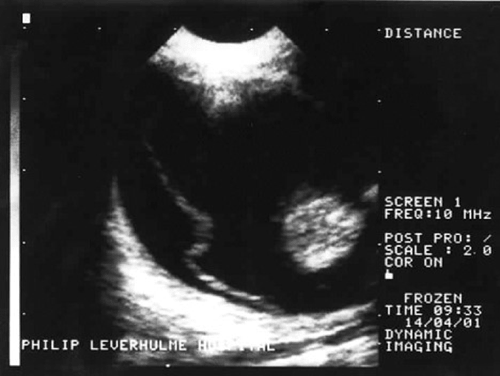
Posterior lens luxation with retinal detachment.
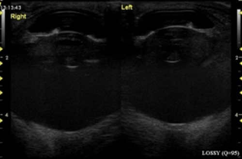
Glaucoma affecting the right eye.
All images kindly provided by Professor Derek Knottenbelt OBE BVM&S DVM&S DipECEIM MRCVS.

