Ultrasonography of the bovine ovary
Ultrasonography of the Bovine Ovary
Ultrasonography of the bovine ovary can be both fascinating and challenging. As the corpus luteum (CL) and ovarian follicles continuously grow and regress, interpretation of ovarian changes can be difficult. Especially as assessment of the ovary can take place at any given point in the ovarian cycle. This article aims to give an overview of the common structures and changes seen when performing ultrasound of the bovine ovaries.
The bovine oestrus cycle lasts 18 to 24 days. During this period, two to three follicular waves develop leading to the emergence of a dominant follicle. Whilst the corpus luteum maintains production of progesterone, the dominant follicles of the initial wave(s) will regress by atresia. Once luteolysis has occurred and progesterone declines, the dominant follicle of the last wave of the cycle may continue to develop and undergo ovulation (Figure 1).
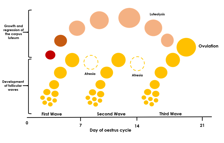
Figure 1. Bovine Oestrus Cycle. This diagram shows the follicular waves and changes of the corpus luteum during one oestrus cycle in which there are three follicular waves.
Follicles
On ultrasound the fluid filled follicles appear as anechoic (black) areas within the ovarian stroma (Figure 2). As follicular waves continuously develop throughout the ovarian cycle, larger follicles >8mm are always present apart from in the first few days of the cycle1(Figure 3). This makes identifying the dominant follicle and predicting ovulation based on follicle size extremely challenging.
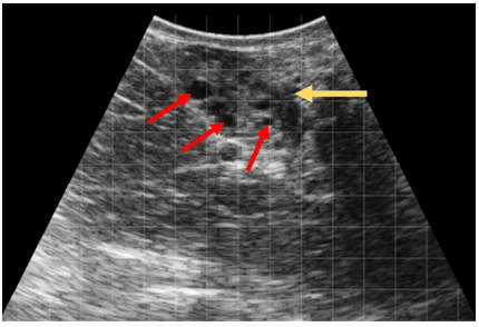
Figure 2. Ovary with small follicles. In this ultrasound image the ovary can be seen (gold arrow) with multiple small follicles (red arrows) within the ovarian stroma.
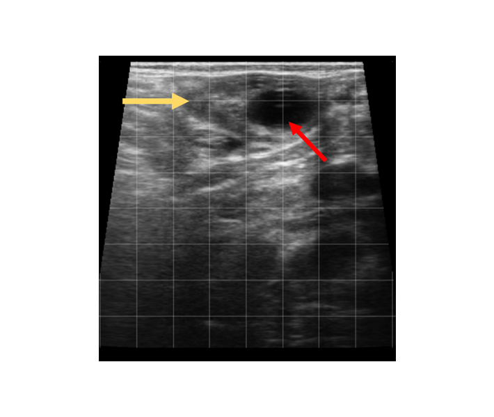
Figure 3. Ovary with a large follicle. In this ultrasound image the ovary (gold arrow) contains a single large (18mm) follicle (red arrow).
Corpus Luteum
On ultrasound the corpus luteum appears as a defined area of hypoechoic tissue within the ovarian stroma (Figure 4.). A central lacuna (fluid-filled cavity) may be seen within a normal CL2 and should not be confused with the presence of a luteal cyst (Figure 5.).
The presence of a CL indicates that oestrus activity has occurred and is a marker of sexual maturity in heifers. The CL may usually be identified on ultrasound examination 4 days after ovulation occurs. If fertilisation of the ovum does not occur and pregnancy is not established, the CL reaches peak size 16 days post-ovulation and then begins to undergo luteolysis.
As the CL persists during pregnancy, localising a CL to the left or right ovary can indicate which uterine horn the embryo will occupy aiding pregnancy diagnosis. Similarly, the presence of multiple CLs (Figure 6.) can aid suspicion of twin pregnancy.
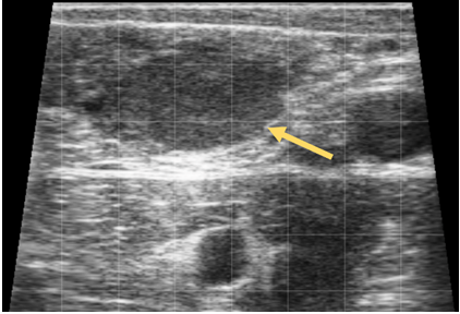
Figure 4. Corpus Luteum. In this image of an ovary, a single CL can be seen (gold arrow).
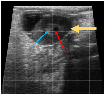
Figure 5. Corpus luteum with central lacuna. This image shows a corpus luteum (gold arrow). There is a central fluid filled lacuna (red arrow) which is partially filled hyperechoic tissue (blue arrow).
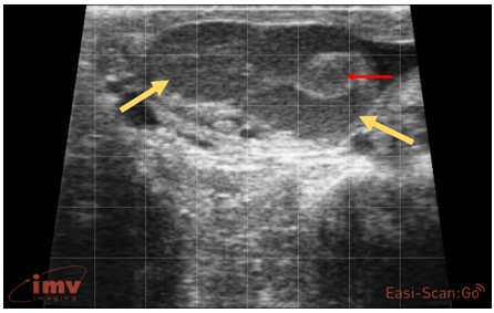
Figure 6. Double CL. In this image two CLs are present on one ovary (gold arrows) indicating a double ovulation has occurred. The right CL has central lacuna which has become filled with echogenic tissue (red arrow).
Ovarian Cysts
Cystic ovarian disease is an important condition to consider, particularly in dairy cattle herd management, as it results in abnormal cyclic activity and a subsequent decrease in fertility. This condition is traditionally defined as the presence of fluid-filled structures greater than 25 mm in diameter on the ovary for longer than 10 days in the absence of a functional CL2. The two types of ovarian cysts resulting in reproductive/ cyclic dysfunction are follicular cysts and luteal cysts. The criteria generally used to define the type of cyst are:
- Follicular cysts – smooth, thin wall (less than 3 mm) (Figure 7.)
- Luteal cysts – thicker wall (greater than 3 mm) due to a lining of luteal tissue (Figure 8.)
However, not all cystic structures adhere strictly to these criteria and differentiating between a luteal cyst and a CL with a large central fluid filled cavity can be challenging.
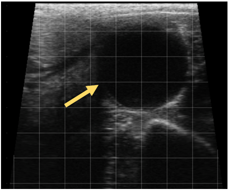
Figure 7. Follicular Cyst. This image shows as large follicular cyst (gold arrow) with an internal diameter of 37mm. Note the thin wall of the cyst.
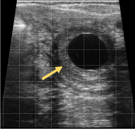
Figure 8. Luteal Cyst. This image shows a luteal cyst (gold arrow) with a central fluid filled area of 25mm. Note the thicker wall of the cyst containing luteal tissue. In some cases, the central fluid filled cavity of luteal cysts may also contain echogenic trabeculae.
Treatment
In addition to allowing early pregnancy diagnosis and the identification of cows that have failed to conceive, ultrasound can guide the use of hormonal therapy and synchronisation programmes.
In the post-partum period, cows that are not showing observed oestrus behaviour can be assessed for the presence of a CL or larger follicles. The absence of both these structures indicate that anovulatory anoestrus is occurring and this allows the selection of appropriate hormonal therapy.
Similarly, the presence of a CL or larger dominant follicle can help inform the practitioner as to whether the cow is in the luteal or follicular phase of the ovarian cycle respectively. This can help select the most appropriate synchronisation protocol to initiate.
Follicular cysts can be treated with gonadotropin-releasing hormone (GnRH) and/or a progesterone releasing device3, luteal cysts will respond to prostaglandin F2α (PGF2α). Modern synchronisation programmes often utilise both PGF2α and GnRH along with progesterone releasing devices and can be used whether there is a luteal cyst or larger cavitary CL making distinguishing between the two structures academic1.
Conclusion
Ultrasonography is extremely rewarding, not only in the assessment of ovarian structures, but in the examination of the bovine uterus and throughout pregnancy as well.
By utilising ultrasound, the practitioner can gain information on ovarian structures and changes that would be impossible with manual palpation alone. This can be very valuable as it informs decision making and targeting any treatment necessary.
References
- Carrière P.D., Gnemmi G., DeCôteaux L., Matsui M., Miyamoto A., Colloton J. (2010) Bovine Ovary. In: Practical Atlas of Ruminant and Camelid Reproductive Ultrasonography, Eds: DeCôteaux L., Gnemmi G., Colloton J. Wiley-Blackwell, Iowa, pp 35-59.
- DeCôteaux L., Gnemmi G., Colloton J. (2009) Ultrasonography of the bovine female genital tract. Veterinary Clinics of North America: Food Animal Practice. 25: 733-752
- Gilbert R.O. (2016) Management of Reproductive Disease in Dairy Cows. Veterinary Clinics of North America: Food Animal Practice 32: 387-410