Practical Approach to Imaging the Equine Stifle
Webinar Details:
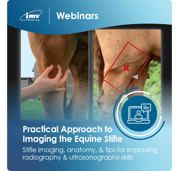
Webinar: Equine Stifle Imaging
The stifle joint is the most complex joint in the equine body and imaging this region can be challenging. The aim of this webinar is to discuss the use of different imaging modalities for evaluation of the stifle, describe the relevant osseous and soft tissue anatomy, and to present a practical approach to radiography and ultrasonography of the region. Filled with helpful hints and tips, this webinar is equally suitable for those wishing to improve their existing imaging skills or confidence, as well as those who perhaps don’t know where to start!
Webinar contents:
- Anatomy overview
- Imaging modalities – what do we know?
- Radiography ‘how-to’, including radiological landmarks
- Ultrasonography ‘how-to’
About the presenter:
Dr Laura Quiney is a Clinical Manager at IMV imaging. Laura qualified from the University of Bristol and subsequently completed an equine orthopaedic internship at the Animal Health Trust (AHT). Following a period working in ambulatory equine practice in Surrey she returned to the AHT as a Junior Clinician, focussing on lameness and poor performance investigation, diagnostic imaging and clinical research. She underwent 4 years of training in radiography, ultrasonography, MRI and scintigraphy under diagnostic imaging specialists, whilst overseeing the equine diagnostic imaging department.