Equine chronic interstitial pneumonia
A case study detailing the symptoms of chronic interstitial pneumonia alongside the use of radiography and ultrasonography. For veterinary use.
A 16-month-old, 300kg Arabian filly presented with a four week history of apparent anhydrosis and generalised loss of body condition despite increased nutritional intake. On clinical examination, the filly was pyrexic (39.0°C), tachycardic (68 beats/min and tachypnoeic (30 breaths/min). Thoracic auscultation revealed the presence of crackles and wheezes diffusely across all lung fields bilaterally. The filly also displayed a marked increase in respiratory effort with a significant abdominal component.

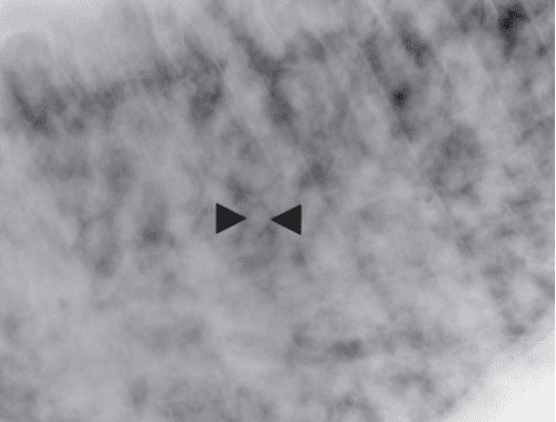
Fig. 1: Lateral radiographic view of the dorsal midthorax. A generalised,
nodular interstitial pattern is evident. Indistinct individual nodules may be
identified (arrowheads), however these appear to be coalescing

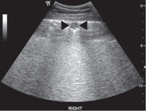
Fig. 2: Transverse ultrasound image of the lungs. A focal, 1-cm diameter,
hypoechoic, subpleural nodule is visible (arrowheads)
Thoracic radiography
The left lateral thoracic radiograph is characterised by a severe, generalised, coalescing, nodular interstitial pattern.
Thoracic ultrasonography
Ultrasound examination of the thorax revealed the presence of numerous hypoechoic nodules deep to the visceral pleura.
On the basis of the history, clinical examination and diagnostic imaging findings, a diagnosis of severe interstitial pneumonia was made. The owner declined further diagnostics and treatment and opted for euthanasia.
Post-mortem findings
On post-mortem examination, multiple, cream coloured, subpleural nodules were visible on gross visual examination of all lung lobes. The cut surface of the lung revealed multiple, spherical, well-circumscribed, firm nodules distributed throughout the pulmonary parenchyma.
Histopathological examination of multiple lung sections confirmed the diagnosis of chronic, diffuse interstitial pneumonia. No causative organism was identified histologically and culture of swabs taken during post-mortem examination failed to identify any significant pathological organism.
Successful treatment of chronic interstitial pneumonia in the foal with broad-spectrum antibiotics and corticosteroids has been previously described. However, adult horses affected with chronic interstitial pneumonia typically respond poorly to treatment due to the presence of secondary fibrosis.
This condition has been termed equine multinodular pulmonary fibrosis (EMPF) and is thought to be associated with equine herpesvirus type 5 infection.

References
Palgrave KA, Palgrave CJ, Rhoads WS and Voges AK. What is your diagnosis? Interstitial pneumonia.J Am Vet Med Assoc. 2008 Sep 1;233(5):711-2
Wong DM, Belgrave RL, Williams KJ, Del Piero F, Alcott CJ, Bolin SR, Marr CM, Nolen-Walston R, Myers RK and Wilkins PA.
Multinodular pulmonary fibrosis in five horses. J Am Vet Med Assoc. 2008 Mar 15;232(6):898-905
