Teat ultrasonography
Check out the Teat ultrasonography
Abnormalities of the teat can have a significant effect on the productivity of individual animals within a herd/flock.
Abnormalities of the teat can have a significant effect on the productivity of individual animals within a herd/flock. In particular, stenosis of the teat can result in a reduction in milk flow impeding efficient milking and also a greater susceptibility to the development of mastitis.
How to scan-
As the teat is not a rigid structure, the ultrasound probe should be applied in a manner that does not significantly distort the shape of the teat. There are two methods for achieving this:
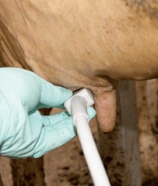
1) Apply the ultrasound probe directly to the teat surface. When imaging the teat/gland junction, an appropriate degree of pressure should be applied to achieve adequate contact but not distort the teat shape.
2) Utilize a ‘standoff’. This technique may improve visualization of the
most superficial region of the teat (directly below the surface of the probe) and the tip of the teat. Ultrasound coupling gel should be applied to all surfaces to improve contact between the probe and ‘standoff’.
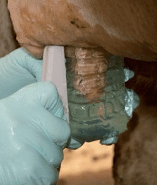
A ‘standoff’, whether purchased or homemade, is an object positioned between the area being scanned and the surface of the probe. It can be a cup or glove filled with water where the teat is submerged.

1 – Scanning with a ‘standoff’

2 – Scanning without a ‘standoff’
Normal anatomy
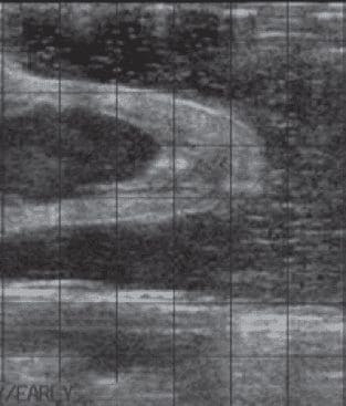
The teat wall is comprised of five layers. Due to their ultrasonographic appearance, they may be grouped into three distinct layers. Milk within the lumen of the normal teat cistern appears anechoic on the ultrasound image. As seen in Figure 3 the three distinct layers are:
1) Inner mucosal layer (teat lining)
2) Middle layer of connective tissue,
muscle, nerves and blood vessels
3) Outer layer of skin.

3 – Ultrasound image of a
normal teat in longitudinal
section (cup of water is being
used as a ‘standoff’)

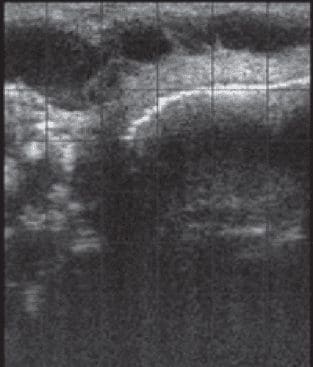
4 – Teat with evidence of fibrosis affecting the lumen
Examples of what you will see:
Nb – these videos include the teat and gland.
Transverse teat without Standoff
Easi-scan normal bovine Teat transverse without standoff from BCF
Longitudinal teat without Standoff
Easi-scan normal bovine teat longitudinal without standoff from BCF
Teat with Standoff
Easi-scan noraml bovine teat with standoff from BCF
Pathology
Figure 4 is an image of a diagnosed case of stenosis/fibrosis of the teat cistern, a common abnormality of the teat. The shape of the teat cistern is distorted and there are hyperechoic strands of material present within the teat cistern, representing fibrosis within the teat lumen. This condition was not grossly apparent externally. Refer to Figure 2 for normal appearance.
Why ultrasound?
Ultrasound may be a useful tool for further investigating the presence and extent of abnormalities affecting the teat. The primary benefit is that various abnormalities of the teat and the teat/gland junction, which can be missed if not grossly evident externally or upon palpation, may be visualized with ultrasound.