Radiography of the equine thoracolumbar spine
If you’ve ever tried to get good radiographs of the equine thoracolumbar spine you will have discovered that it can be quite tricky at times!
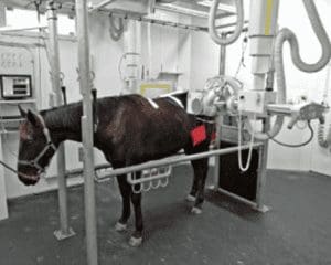
Digital radiography has certainly made life easier, but there remain issues caused by the physical size and shape of the horse in this anatomical location. Tissue thickness at the level of the vertebral bodies can be quite considerable, necessitating the use of very high kV and mAs settings. This often means there’s a need for gantry-mounted generators and wall-mounted cassette holders, and techniques to ensure the patient stands as still as possible also help!
Image courtesy of www.podoblock.com
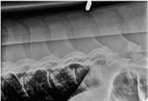
X-ray scatter can be a real issue when radiographing this region in horses and there are several measures that one can take to minimise its negative effects: placement of a lead sheet behind the cassette will help to reduce some of this scatter (also increasing staff safety). Use of a grid with a grid ratio of 10:1 or even 12:1 will improve image quality but will also mean that even higher exposure factors are required, which can be problematic if equipment limitations come into play.
Image courtesy of www.podoblock.com
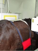
One major issue with radiographing this region is the dramatic difference in the thickness of the soft tissue mass overlying the tips of the dorsal spinous processes (DSPs) compared to the same tissue thickness at the vertebral bodies in lateral radiographs. The result is either underexposed vertebral bodies and overexposed DSPs, or a need to take two radiographs of the same region using different exposure settings. In this instance the use of aluminium filter wedges can compensate for this disparity in tissue thickness, producing a radiograph with a more even exposure over the whole region, obviating the need to take two radiographs.
Image courtesy of www.podoblock.com


