IMV imaging Courses 2026
Small Animal Bronchoscopy and Rigid Endoscopy
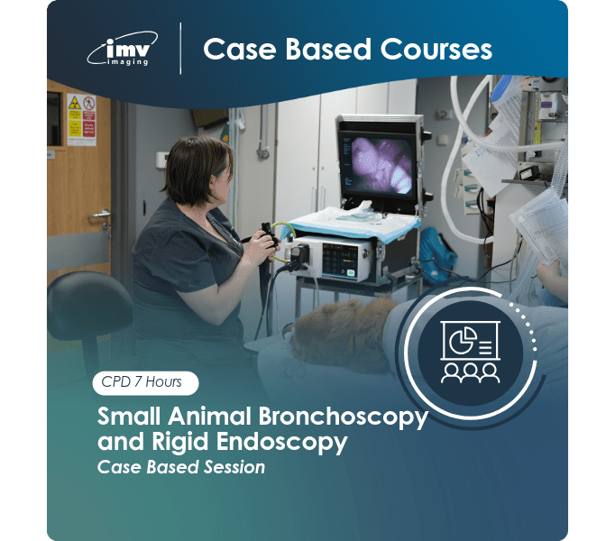
Course Overview:
This one-day course will be a case based course. We will deliver the content through lectures including images and videos to allow delegates to understand the application and the clinical rationale of the use of bronchoscopy and rigid endoscopy in small animals as an aid to work up clinical medical cases.
Topics Covered:
- How to approach bronchoscopy – what clinical presentations or cases would benefit from bronchoscopy and when it would be useful – when to scope and when not to?
- How to practically carry out the procedure in both cats and dogs
- What equipment you need
- How to do a proper BAL in both cats and dogs? What samples you need and how to get them?
- How to investigate nasal disease in both dogs and cats and the role of rhinoscopy – optimising case selection and understanding the limitations of each of the available techniques.
- How to perform cystoscopy with both rigid and flexible equipment – practical step by step guides on how to actually carry out the procedure? When and how to take biopsies?
- Case-based discussions – reviewing and discussing interesting cases where endoscopy has been useful in influencing the outcome.
Equipment and Facilities:
There is no requirement to bring any equipment or patients to this course, given the nature of this training, this will be a theory only course.
The day will start at 9am for Tea/coffee. Lectures to start at 9.30am and the day will finish around 5.30pm. It includes a full 2 course lunch and course notes will be provided.
Location:
IMV Imaging,
Unit 2, Block 3,
City North Business Campus,
Gormanston,
Co.Meath, K32 ER81
Speaker: Mark Dunning MA VetMB CertSAM PhD DipECVIM-CA FRCVS. RCVS and EBVS European Specialist in Small Animal Internal Medicine
CPD: 7 hours CPD. VCI CVE credits pending approval
Cost: £440 + VAT / €520 + VAT – Includes 2 course lunch and course notes
If you have any further questions, please contact fiona.mcfarland@imv-imaging.com / 0044 7760 908463
IMV imaging Courses 2026
Learning has always been at the heart of IMV imaging. For three decades we have been running practical ‘hands-on’ ultrasound training courses and we are delighted to also offer radiography courses for vet nurses. We are committed to ensuring you can make the most of your equipment.
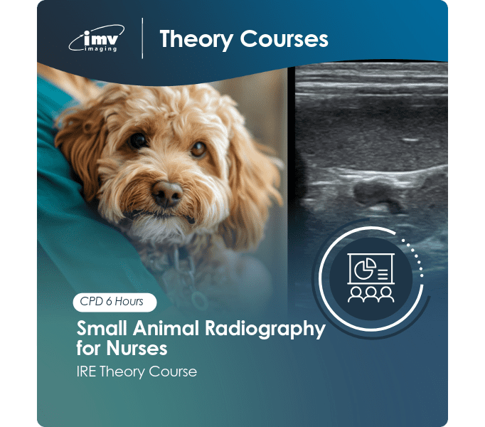
Small Animal Radiology for Nurses
Course Overview:
This one day course is open to all members of the veterinary nursing team, both Registered Veterinary Nurses and Student Veterinary nurses. It will be of benefit to anyone with an interest in radiography and whom want to improve their skills and knowledge in this area.
This course will provide the theoretical and practical tools to give participants the confidence to complete excellent radiographs covering both abdominal and thoracic studies along with MSK and joint approaches. It covers a systematic approach to understanding the physics of radiography, how to position correctly, and how to avoid the common pitfalls. During the session we will focus on practical tips and hints. This is a very interactive day full of lots of case discussion with quizzes and will allow you to ask our expert all the questions you have had about all things radiography related. This day has been thoroughly enjoyed by previous delegates.
We aim to meet at 9.30am for Tea/coffee and lectures will start at 10.00am. It includes a full 2 course lunch and course notes will be provided.
Topics Covered:
- Radiographic physics and understanding your equipment – what does KV and mAs mean, how to choose exposures and take a good radiograph
- Positioning and radiographic anatomy – top tips and how to maximise your time taking radiographs covering all body areas – thorax, abdomen and musculoskeletal radiography
- Exposures and artefacts – how to tell when the image is diagnostic, what to do or change when things are wrong, common pitfalls, mistakes and artefacts and how to avoid them
- Recognising Pathology – what does normal look like and how to pick up the abnormal including case studies
- Full interactive session on reading radiographs and how to get the best out of your skills
Equipment and Facilities: The day will be informal and relaxed with maximum time for hands-on learning. We aim to meet at 9.30am for Tea/coffee and lectures will start at 10.00am. 2 course lunch will be provided and course notes will be provided.
Location:
IMV Imaging,
Unit 2, Block 3,
City North Business Campus,
Gormanston,
Co.Meath, K32 ER81
Speaker: Mark Coia BVMS DipECVDI MRCVS RCVS and EBVS European Specialist in Diagnostic Imaging
CPD: 6 hours CPD. VCI CVE credits pending approval
Cost: £250 + VAT / €300 + VAT – Includes 2 course lunch and course notes
If you have any further questions, please contact fiona.mcfarland@imv-imaging.com / 0044 7760 908463
Ultrasound Course 2026
Learning has always been at the heart of IMV imaging. For three decades we have been running practical ‘hands-on’ ultrasound training courses and we are now delighted to also able to offer courses for vet nurses. We are committed to ensuring you can make the most of your equipment.
Nurse Ultrasound Course
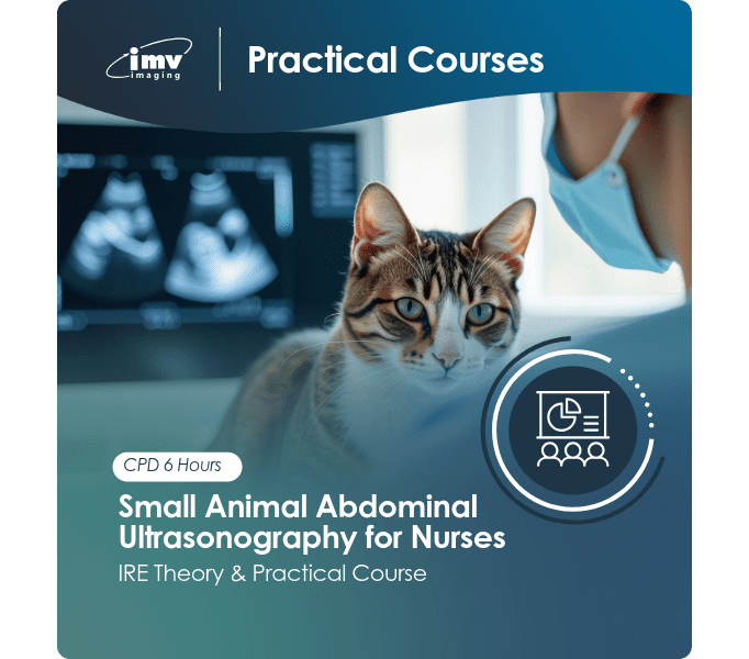
Course Overview: Ultrasound! – Can Veterinary Nurses Scan? Yes they Can!
This combined theory and practical course is aimed at both absolute beginners and those with ultrasound skills already in development, we will guide you through everything you need to know to get to grips with abdominal ultrasonography. From patient preparation, machine controls and how ultrasound is applied, we will build your skills and get you confidently performing both point of care ultrasound examinations and identifying all abdominal organs.
Theory will also include case discussion and it will be a very interactive session, including assessing images and videos, watching a full demonstration of an abdominal scan and then taking part in practical sessions in a relaxed, friendly and informal environment.
Topics Covered:
- Machine setup and patient preparation
- How ultrasound artefacts appear when scanning, how they can be useful to you or how to avoid them!
- Machine controls, what they mean and how to optimise the image
- How to identify all the major abdominal organs – Liver, Gall bladder, GIT, kidneys, spleen, major vessels and lymph nodes
- What significant pathology looks like and when to be concerned
- How nurses can apply ultrasound techniques to case management
- What to look out for in an emergency case presentation including APOCUS techniques
- Guidance and in-practice experiences from our in house vet Fiona
Equipment and Facilities:
We ask that the majority of delegates bring a dog to scan to allow enough hands on practice, so please let us know if you cannot bring a patient before booking. It is important that the dogs can be clipped as required for an abdominal examination.
Kennelling accommodation for the dogs will be available to use if required. Various ultrasound machines will be available to use on the day, however, if you want to bring your own for practice you are welcome to do so, just let us know.
The day will be informal and relaxed with maximum time for hands-on learning. Arrive for tea and coffee from 9am, ready to begin the lectures at 9.30am. We will aim to finish by 5.30pm.
Places are limited to a maximum of 12 delegates so book your place early to avoid disappointment. It includes a full 2 course lunch and course notes will be provided.
Location:
- Fri 1st May – CAFRE Greenmount Campus, 45 Tirgracy Rd, Antrim, Co.Antrim BT41 4PS
- Fri 4th September – IMV Imaging, Unit 2, Block 3, City North Business Campus, Gormanston, Co.Meath K32 ER81
- November – Date & venueTBC (Cork area)
Speaker: Fiona McFarland BVSc MRCVS – Small Animal Veterinary Surgeon and Clinical Sales Manager IMV Imaging
CPD – 6 hours CPD
Cost – £250 + VAT / €300 + VAT – Includes 2 course lunch and course notes
If you have any further questions, please contact fiona.mcfarland@imv-imaging.com / 0044 7760 908463
Small Animal Endoscopy Course 2026
Learning has always been at the heart of IMV imaging. For three decades we have been running practical ‘hands-on’ ultrasound training courses and we are now delighted to also able to offer endoscopy courses. We are committed to ensuring you can make the most of your endoscopy equipment.
Small Animal Endoscopy Course – Exploring the Gastrointestinal Tract – from FBs to Biopsies!

Course Overview:
This one-day course will be delivered as a mixture of theory and practical learning focussing on the use of flexible endoscopy within the gastrointestinal tract of dogs and cats. We will get you set to tackle all sorts of GIT endoscopy, and not only ensure you know how to drive an endoscope but get you ready for retrieving foreign bodies (including all the tips and tricks to this), but also leave you feeling confident taking biopsies with advice on when and how to do it. Practical techniques will be demonstrated by Mark and practiced by delegates using a number of models including our canine mannequins. It’s like real life endoscopy!
Topics Covered:
- Understanding the basic principles of endoscopy
- Understanding the strengths and limitations of the technique as an aid to diagnosis, when is endoscopy a good next step?
- How to use the equipment, how to manipulate and direct flexible endoscopes – get ready to learn how to drive this machine!
- How to navigate the gastrointestinal tract, including the dreaded pylorus, Mark will give you all the tips you need to get through the tricky places
- Recognising pathology and then when and how to take good biopsies throughout the GIT
- Techniques and equipment required to remove gastric foreign bodies – get those tricky bits out safely
- Placement of PEG tubes (why, when and how to do it)
Equipment, Facilities and Agenda:
There is no requirement to bring any equipment or patients to this course, we will provide everything.
The day will start at 9am for Tea/coffee. Lectures to start at 9.30am with a mixture of lectures and practical scanning sessions in both the morning and afternoon sessions. The day will finish around 5.30pm, It includes a full 2 course lunch and course notes will be provided.
Location:
IMV Imaging Ireland Office
Unit 2, Block 3,
City North Business Campus,
Gormanston, Co. Meath,
K32 ER81
Speaker: Professor Mark Dunning MA VetMB PhD CertSAM DipECVIM-SA FRCVS
CPD: 7 hours CPD , VCI credits pending approval
Cost – £440 + VAT / €520 + VAT – includes lunch and course notes.
If you have any further questions, please contact fiona.mcfarland@imv-imaging.com / 0044 7760 908463
IMV imaging Courses 2026
‘Maximising your full potential – scan your way to success!’ Theory & Case Discussion
Advanced Abdominal Ultrasonography
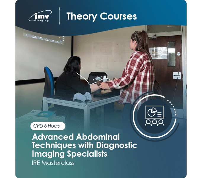
Course Overview:
This advanced veterinary ultrasound session is a theoretical and case-based interactive session, designed for clinicians with the intention to follow on from the beginners and intermediate ultrasound courses; allowing for you to take your skills to the next level and ‘scan your way to success’ equipping you with the knowledge needed to help with clinical reasoning, decision making and action.
Are you a small animal clinician with a real interest in veterinary ultrasound and have already got to grips with the basics? You feel competent and confident finding the vital organs within the abdomen and appreciate normal variations and you now want to learn what the pathology means? What am I looking at when it goes wrong? If so, this one day-course sounds ideal to help with your journey when exploring abdominal ultrasound to an advanced level.
During the course we will discuss abnormal findings, correlation with other imaging techniques (namely radiography), discuss a variety of ultrasound cases relating to various body organs, how to formulate a differential list, approaching a paradigm of formulating a structured report, covering the entire abdomen but with a special focus on systematic approach of the gastrointestinal tract, the adrenals, abdominal lymph nodes as well as the pancreas; the session will allow the opportunity to ask all the questions you like as it’s very interactive.
Topics Covered:
- Ultrasonography of the emergency patient
- Approach to Abdominal Point of Care Ultrasound (A-FAST) Focused assessment of sonography for trauma, detecting and excluding visceral injuries including Peritoneal and pleural effusion
- Theoretical overview of the liver, gastrointestinal tract, abdominal lymph nodes, pancreas, adrenals, urogenital tract
- Approaching abnormalities and what do these possibly indicate
- Clinical decision making and producing reports. Case based discussion of the various body systems
- Approach to the gastrointestinal tract and feeling comfortable with a thorough review. Ultrasonographic approach to the vomiting patient, how likely am I to find pathology? Evaluating causes of diarrhoea or GIT bleeding. Helping with decision to ‘cut or not to cut’?
- Advanced doppler ultrasound and abdominal vessels: Portal vessel and mesenteric vessels, hepatic vasculature, aorta, and caudal vena cava
- Approach to Ultrasound guided tissue sampling – when is sampling indicated?
PLEASE NOTE – As this course is covering pathology and case discussion, it is a theory only course and no need to bring animals for practical scanning.
Location:
IMV imaging Unit 2, Block 3
City North Business Campus
Gormanston, Co. Meath
K32 ER81
Times – 9.30am – 5.30pm
Speaker: Mark Coia BVMS DipECVDI MRCVS RCVS and EBVS European Specialist in Diagnostic Imaging
CPD: 6.0 hours CPD. VCI CVE credits pending approval
Cost: £400 + VAT / €460 + VAT – Includes 2 course lunch and course notes
If you have any further questions, please contact fiona.mcfarland@imv-imaging.com / 0044 7760 908463
IMV imaging Courses 2026
Small Animal Radiography
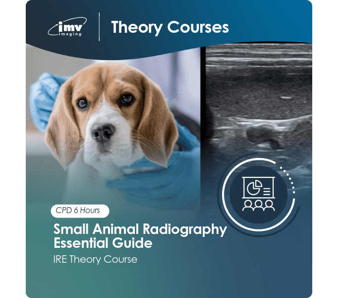
Course Overview:
This one day course is suitable for all Small Animal clinicians and will provide the theoretical and practical tools to give participants the confidence to examine and interpret straightforward and complex radiographic studies of cases which are commonly encountered. It covers a systematic approach to the radiological interpretation of all organ systems in the thoracic and abdominal cavities while discussing several orthopaedic conditions also. During the session we will focus on how to recognise normal and abnormal findings as well touching on the common indications for the use of contrast studies for the evaluation of the gastrointestinal and urinary tract.
The course aims to help improve competency and confidence while assisting participants with discerning the information on radiographs that is most useful for case management. Practical film reading session will be included throughout the day. This is a very interactive day full of lots of case discussion, thoroughly enjoyed by previous delegates.
We aim to meet at 9am for Tea/coffee and lectures to start at 9.30am. It includes a full 2 course lunch and course notes will be provided.
Topics Covered:
- Understand standard radiographic views and the benefit of ancillary projections to assist with investigation
- Apply knowledge to perform an interpretation of various imaging modalities
- Discuss the uses of radiography in the investigation of diseases
- Understand how the different diagnostic imaging modalities can be used in their diagnosis
- Discuss benefits and limitations of various aspects of Small Animal Radiology
- Develop an effective and practical technique for reading radiographs of dogs and cats relating to the thorax, abdomen and musculoskeletal system
- Recognise and describe abnormalities seen on diagnostic images and associate these with the underlying pathophysiology
- Recognition of normal versus abnormal findings when evaluating radiographs, interpret findings and gain confidence with approaching basic and complex thoracic, abdominal and musculoskeletal radiograph
- Clinical reasoning: extract the information on these radiographs which is useful for patient management and determining outcomes
- Theoretical and practical film reading sessions: Practice your radiograph reading skills using a large number of radiographic cases
- Myth vs Truth: Debunking myths- this is your chance to ask any questions you may have and put any myths to bed
Equipment and Facilities: The day will be informal and relaxed with maximum time for hands-on learning. Arrive for tea and coffee from 9.00am, ready to begin the lectures at 9.30am. 2 course lunch will be provided. We will aim to finish by 5.30pm.
Location:
IMV Imaging,
Unit 2, Block 3,
City North Business Campus,
Gormanston,
Co.Meath, K32 ER81
Speaker: Mark Coia BVMS DipECVDI MRCVS RCVS and EBVS European Specialist in Diagnostic Imaging
CPD: 6 hours CPD. VCI CVE credits pending approval
Cost: £350 + VAT / €410 + VAT – Includes 2 course lunch and course notes
If you have any further questions, please contact fiona.mcfarland@imv-imaging.com / 0044 7760 908463
IMV imaging Courses 2026
‘Logic will get you from Adrenals to Bladder and the whole way through the Abdomen’
Abdo Plus! – Next steps to abdominal ultrasonography
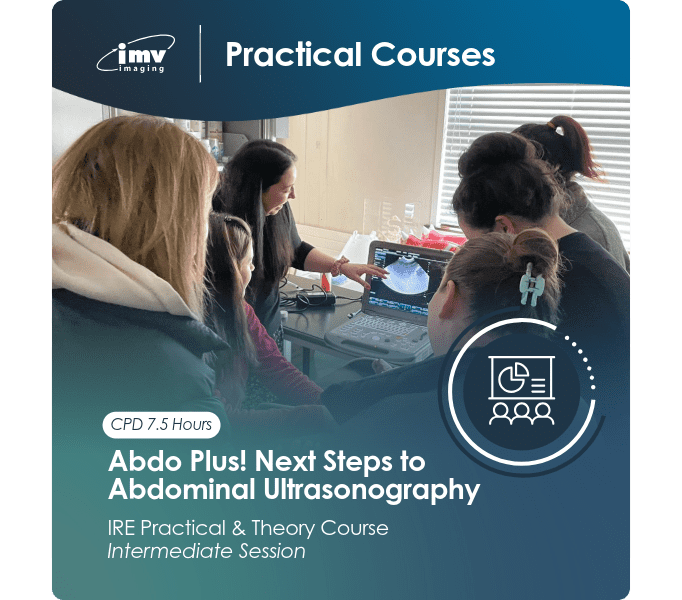
Course Overview:
This full day course is aimed at veterinary surgeons comfortable with basic abdominal ultrasonography and who want to take their skills a step further, tackling trickier things. With a mixture of lectures, demonstrations and practical hands-on sessions, you will soon get to grips with the entire tour of the abdomen.
Throughout the course you will learn the normal and pathological appearance of all major organs along with adrenal glands, pancreas, major vessels and abdominal lymph nodes. You will learn the landmarks to guide you to the subtler structures and you will examine the anatomy of the abdominal vasculature with the use of doppler ultrasonography.
Topics Covered:
- Get comfortable with the GI tract – learn to find your way around the stomach, small intestine, including duodenum, ileum, ICCJ and colon
- Look further at kidneys, the urinary and reproductive tracts and then where to find the adrenal glands and what to look out for
- Learn the landmarks that help us to locate the pancreas and what to expect
- Find and examine the major blood vessels and abdominal lymph nodes
- Understanding the use of doppler ultrasonography within the abdomen
- Put it into practice – get hands-on!
Equipment and Facilities:
We ask that the majority of delegates bring a dog to scan on the day, please let us know if you cannot bring a patient before booking. It is important that the dogs can be clipped as required for a full abdominal scan.
Kennelling accommodation for the dogs will be available to use if required. Various ultrasound machines will be available to use on the day, however, if you want to bring your own for practice you are welcome to do so, just let us know.
The day will be informal and relaxed with maximum time for hands-on learning. Arrive for tea and coffee from 8.30am, ready to begin the lectures at 9am. We will aim to finish by 5.30pm. Places are limited to a maximum of 10 delegates so book your place early to avoid disappointment. Course notes will be provided.
Location:
IMV imaging Unit 2, Block 3
City North Business Campus
Gormanston, Co. Meath
K32 ER81
CPD: 7.5 hours CPD. VCI CVE credits pending approval
Cost: £540 + VAT / €640 + VAT – Includes 2 course lunch and course notes
If you have any further questions, please contact fiona.mcfarland@imv-imaging.com / 0044 7760 908463
IMV imaging Courses 2026
Finding your way around ‘The Thorax and the Abdomen’
Abdo-Cardabra! Small Animal Basic Ultrasound
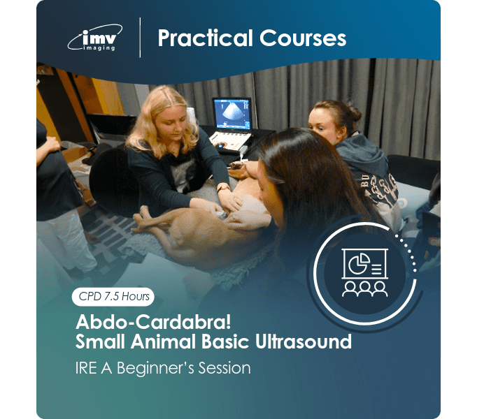
Course Overview:
This full day course is aimed at vets with minimal experience of diagnostic ultrasound and those who feel that they could get much more from their ultrasound machine for both basic abdominal ultrasonography and basic echocardiography.
Via a blend of lectures, demonstrations, and practical hands-on sessions you will gain the fundamental knowledge to improve your skills or get started if you don’t know where to begin.
Topics Covered:
- Basic ultrasound physics – understand how an image is formed and what to do when things are not as they appear
- Get familiar with the machine – learn the core controls and how to get the best image, making your image better when you perform an ultrasound scan, depending on what you are looking at
- Indications – when to use ultrasound examinations – when is it a useful diagnostic tool?
- A logical approach to the main organs of the abdomen and a basic approach to the heart
- What normal looks like – Liver, gall bladder, spleen, GI Tract, kidneys, urogenital tracts and the general approach to assessing the abdomen
- Learn the normal appearance of the cardiac anatomy including normal right sided echocardiographic 2D views and their interpretation
- Put it into practice – get stuck in!
Equipment and Facilities:
We ask that most delegates bring a dog to scan, but it is not essential. Please let us know if you cannot bring a patient before booking. It is important that the dogs can be clipped for full abdominal and echocardiographic examinations.
Kennelling accommodation for the dogs will be available to use if required. Various ultrasound machines will be available to use on the day, however, if you want to bring your own for practice you are welcome to do so, just let us know. The day will be informal and relaxed with maximum time for hands-on learning.
Arrive for tea and coffee from 8.30am, ready to begin the lectures at 9am. We will aim to finish by 5.30pm. Places are limited to a maximum of 10 delegates so book your place early to avoid disappointment. Course notes will be provided
Location:
IMV imaging Unit 2, Block 3
City North Business Campus
Gormanston, Co. Meath
K32 ER81
Speaker: Mark Coia BVMS DipECVDI MRCVS RCVS and EBVS European Specialist in Diagnostic Imaging
CPD: 7.5 hours CPD. VCI CVE credits pending approval
Cost: £520 + VAT / €620 + VAT – Includes 2 course lunch and course notes
If you have any further questions, please contact fiona.mcfarland@imv-imaging.com / 0044 7760 908463
Looking for something more advanced?

IMV imaging Courses 2026
ECHO Plus – Intermediate Small Animal Echocardiography
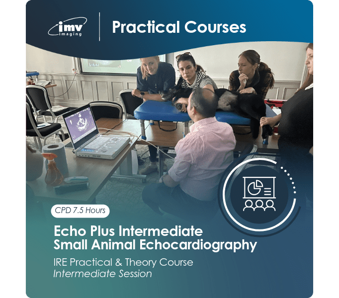
Course Overview:
This full day course is for veterinary surgeons comfortable with basic echocardiography and who want to refresh the basics and learn more. The day will be a mixture of interactive lectures, critical appraisal of echo images and hands-on scanning time. Delegates should be comfortable with very basic right sided views, however we will cover a brief refresher and spend some time perfecting these images, then learn to use colour and spectral doppler as well as working on left sided views.
Topics Covered:
- A refresher of basic 2D right-sided echo views and how to optimise them
- An introduction to Doppler ultrasonography – how, when and why it is used
- How to perform the left-sided echocardiographic views
- Get to grips with the measurements needed to make diagnoses and those we can use to assess cardiac function in more detail
- Spotting pathological changes
- Put it into practice – get stuck in!
Equipment and Facilities:
We ask that the majority of delegates bring a dog to scan to allow enough hands on practice, so please let us know if you cannot bring a patient before booking. It is important that the dogs can be clipped as required for an echocardiographic examination.
Kennelling accommodation for the dogs will be available to use if required. Various ultrasound machines will be available to use on the day, however, if you want to bring your own for practice you are welcome to do so, just let us know.
The day will be informal and relaxed with maximum time for hands-on learning. Arrive for tea and coffee from 8.30am, ready to begin the lectures at 9am. We will aim to finish by 5.30pm. Places are limited to a maximum of 8 delegates so book your place early to avoid disappointment. Course notes will be provided
Location:
IMV imaging Unit 2, Block 3
City North Business Campus
Gormanston, Co. Meath
K32 ER81
Speaker: Chris Linney BVSc GPCertSAP CertAVP(VC) DipECVIMCA (Cardiology) MRCVS
RCVS and EBVS Specialist in Small Animal Cardiology
CPD: 7.5 hours CPD. VCI CVE credits pending approval
Cost – Intermediate Courses: £540 + VAT / €640 + VAT – Includes 2 course lunch and course notes
If you have any further questions, please contact fiona.mcfarland@imv-imaging.com / 0044 7760 908463
IMV imaging Courses 2026
MOO-ving on: the next step in bovine ultrasonography
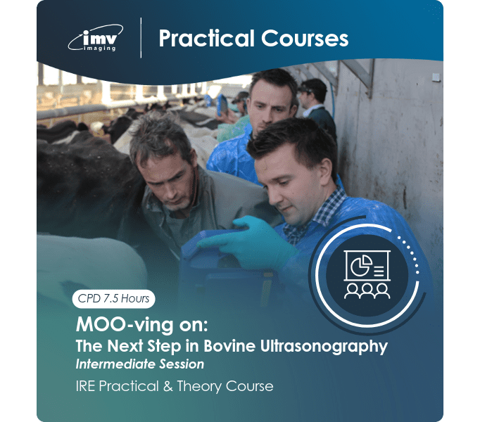
Course Overview: This course is aimed at those who are already comfortable with basic pregnancy diagnosis via ultrasound and want to start using ultrasound for more pre breeding fertility work and advancing their staging of pregnancy and sexing embryos.
Are you currently using ultrasound for your bovine pregnancy checks? If so, can you advise the farmer to expect a bull or a heifer calf? Can you tell the difference between the ultrasound appearance of follicular and luteal cysts? Do you want to be able to estimate the stage of pregnancy more accurately?
This course is more focussed on theory and case discussion crush side. We assume that you have either already done the basic course or are happy with basic pregnancy diagnosis. The course will give you an opportunity to talk through cases as they come up the race on one of Ireland’s largest and most progressive dairy farms. There will be a huge selection of cows to work on, and we will talk through each case as a group.
Topics Covered:
- Accurate staging/ageing of the pregnancy – refresh your knowledge and get those days accurate
- The pre breeding non pregnant cow – Follicular wave assessment, identifying individual cow’s cycles and how to apply practical hormonal and therapeutic management to get her back in calf as quickly as possible
- Detection of twins – its importance and benefit to the farmer
- Foetal sexing – probe positioning, tips and tricks and making the right call on bull or heifer
- Cystic ovarian disease – follicular or luteal?
- Endometritis, pyometra and unusual ultrasound images – when does it look wrong?
- Case evaluation of ovarian and uterine problems including advice on treatment protocols
Agenda: 8.30 – Arrival, Tea/Coffee. The course will start at 9am with a mixture of lectures and practical scanning sessions in both the morning and afternoon sessions. The day will finish around 5.30pm.
Equipment and Facilities: Please bring full clean PPE and gloves/gel for the practical scanning portion of the day. All ultrasound equipment will be provided on the day
Location:
Bingham’s Farm
203 Sevenmile Straight
Antrim, Co. Antrim
BT29 4YR
Speaker: John Dawson, BVMS CERT. CHP MRCVS
CPD: 7.5 hours CPD. VCI CVE points pending approval
Cost: £500 + VAT / €600 + VAT – CPD course notes provided
If you have any further questions, please contact fiona.mcfarland@imv-imaging.com / 0044 7760 908463